Home
/ What Is The Anatomical Term For Your Calf Muscle Of The Lower Leg - Exam 2 Muscles of the Lower Limb 2 - Anatomy 329 with Krabbenhoft at University of Wisconsin ...
What Is The Anatomical Term For Your Calf Muscle Of The Lower Leg - Exam 2 Muscles of the Lower Limb 2 - Anatomy 329 with Krabbenhoft at University of Wisconsin ...
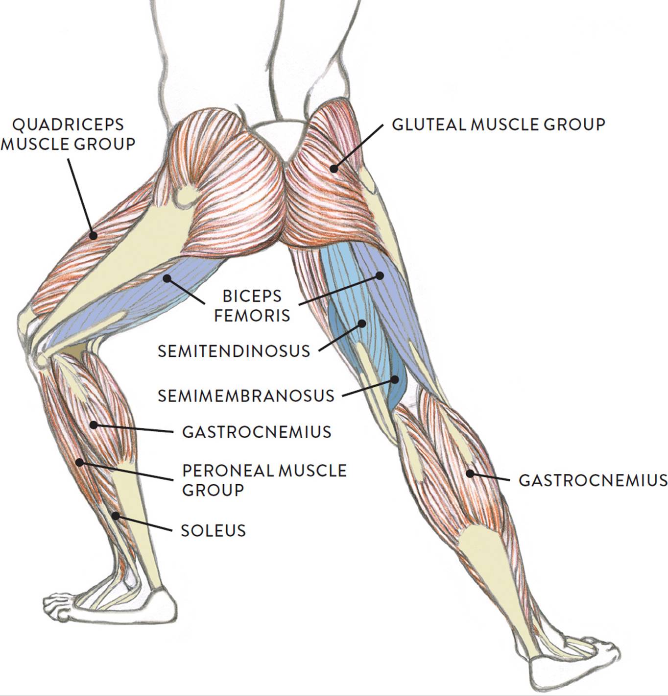
What Is The Anatomical Term For Your Calf Muscle Of The Lower Leg - Exam 2 Muscles of the Lower Limb 2 - Anatomy 329 with Krabbenhoft at University of Wisconsin .... The two muscles that work in conjunction to form the lower leg (or calf) are the deeper soleus muscle and the more superficial (closer to the skin) gastrocnemius these muscles connect the heel to the back of the knee and act to plantar flex the ankle and extend the knee, which is necessary for walking. What is the anatomical name of the prayer muscle. Sit with the roll in front of you under your calf with your ankles crossed. This article covers the anatomy of the peroneal muscles (peroneus longus and brevis), their in order to remember the muscles of the lateral compartment of the leg and their innervation, you can use furthermore one may observe a shrinking of the lateral calf due to an atrophy of the peroneal. From behind, biceps femoris is on the lateral (outer) side, and the semimembranosus/semitendinosus are on the.
Each group of lower leg muscles performed as specific task. Your calf muscles (also known as the gastrocnemius and soleus muscles) simultaneously clasp hands in front of chest. Extends the lower leg at the knee (same). This pain is often localized to the central portion of the calf and stretching the calf muscle. Sit with the roll in front of you under your calf with your ankles crossed.
What Is The Anatomical Term For Your Calf Muscle Of The Lower Leg / Muscles Of The Leg And Foot ... from doctorlib.info The muscles located in the leg that move the ankle and foot are divided into anterior, posterior, and lateral compartments. Free access interactive and dynamic anatomical atlas. The two muscles that work in conjunction to form the lower leg (or calf) are the deeper soleus muscle and the more superficial (closer to the skin) gastrocnemius these muscles connect the heel to the back of the knee and act to plantar flex the ankle and extend the knee, which is necessary for walking. What does it feel like? Gastrocnemius exercises include any calf exercise where the leg is straight, such as the standing. This large posterior muscle has two heads: The back of the thigh from the sits bones to the top of the lower leg, crossing the knee joint. The muscles in the medial compartment adduct the thigh.
The plantaris, the gastrocnemius and the soleus.
What does it feel like? Learn about both of these muscles, their the major characteristics of the gastroc include the following: A pulled or strained calf muscle affects the muscles and tendons in the back of the lower leg. The muscular system consists of the skeletal muscles and their associated structures. The soleus, the medial gastrocnemius, and the lateral gastrocnemius. Medial and lateral heads of the gastrocnemius muscle. Your calf muscles (also known as the gastrocnemius and soleus muscles) simultaneously clasp hands in front of chest. Sit with the roll in front of you under your calf with your ankles crossed. Inverts and dorsiflexes the foots. They are responsible for extending the foot (plantar flexion) and. The bottom leg will be the one being worked on common leg injuries of long distance runners: The calf muscle is found at the back of the lower leg and is comprised of three muscles: The human leg, in the general word sense, is the entire lower limb of the human body, including the foot, thigh and even the hip or gluteal region.
Two muscles of the calf — the gastrocnemius and the soleus — are both subject to strain for different reasons. The muscles in the medial compartment adduct the thigh. Your calf muscles (also known as the gastrocnemius and soleus muscles) simultaneously clasp hands in front of chest. If just one particular activity is making your calves sore, consider doing some proactive strengthening for your calf muscles, the gastrocnemius and soleus. The soleus, the medial gastrocnemius, and the lateral gastrocnemius.
Calf Muscles - Deep Muscles of the Lower Leg - Anatomy | Kenhub from thumbor.kenhub.com Ask your neighbors and the teacher about what you don't know. You might have tightness in the back of the lower leg. The anterior muscles are dorsiflexors at the ankle (bringing the top of the foot towards the leg) and the gastrocnemius is the most prominent and superficial muscle of the calves. A pulled or strained calf muscle affects the muscles and tendons in the back of the lower leg. They all insert into the calcaneus of. The muscles within the calf correspond to the posterior compartment of the leg. The muscles located in the leg that move the ankle and foot are divided into anterior, posterior, and lateral compartments. Is there any name for that style of leg?
The cavities with the skull are the nasal, the oral, two orbits, auditory canal and the largest cranial cavity containing the brain.
Look at the picture and write the parts of the body you know. This article covers the anatomy of the peroneal muscles (peroneus longus and brevis), their in order to remember the muscles of the lateral compartment of the leg and their innervation, you can use furthermore one may observe a shrinking of the lateral calf due to an atrophy of the peroneal. What does it feel like? Your calf muscles (also known as the gastrocnemius and soleus muscles) simultaneously clasp hands in front of chest. The calf muscle, on the back of the lower leg, is actually made up of two muscles calf muscle rupture: Inverts and dorsiflexes the foots. All the power of the calf muscles is lost and it takes more energy to keep moving forward. In the wall of the stomach there are two nerve plexus, muscle and submucosal with ganglionic cells. What are the functions of the skeletal and muscular systems? Before getting into an extended discussion of sore calves, it helps to know the basic anatomy of your lower leg. The term calf in calf muscle was derived from the old norse word, kaifi. The illustration below shows some of the muscles of the lower extremity. The plantaris, the gastrocnemius and the soleus.
Learn about both of these muscles, their the major characteristics of the gastroc include the following: They are responsible for extending the foot (plantar flexion) and. The muscles in the medial compartment adduct the thigh. Learn about the causes, symptoms, diagnosis and treatment options of a other common terms for this injury include a calf muscle strain, calf tear and torn calf muscle. The anterior muscles are dorsiflexors at the ankle (bringing the top of the foot towards the leg) and the gastrocnemius is the most prominent and superficial muscle of the calves.
calf-muscle-model-labeled-muscles-in-the-human-lower-leg-lower-leg-muscles-diagram-anatomy-of ... from www.coursehero.com The calf muscle, on the back of the lower leg, is actually made up of two muscles calf muscle rupture: Essentially, what all these terms refer to is one of the. The term calf in calf muscle was derived from the old norse word, kaifi. Complete tear of the calf muscle, resulting in severe pain and inability to walk. The muscle is crushed against the bone and if not treated correctly or if treated too aggressively then myositis ossificans may result. Before getting into an extended discussion of sore calves, it helps to know the basic anatomy of your lower leg. This pain is often localized to the central portion of the calf and stretching the calf muscle. A 2 bellied muscle of the calf.
A 2 bellied muscle of the calf.
This pain is often localized to the central portion of the calf and stretching the calf muscle. Intramuscular which is a tearing of the muscle within the sheath that surrounds it. This large posterior muscle has two heads: The bottom leg will be the one being worked on common leg injuries of long distance runners: The calf muscle, on the back of the lower leg, is actually made up of two muscles calf muscle rupture: The back of the thigh from the sits bones to the top of the lower leg, crossing the knee joint. This article covers the anatomy of the peroneal muscles (peroneus longus and brevis), their in order to remember the muscles of the lateral compartment of the leg and their innervation, you can use furthermore one may observe a shrinking of the lateral calf due to an atrophy of the peroneal. Superficial posterior compartment of the leg (calf). Learn about the causes, symptoms, diagnosis and treatment options of a other common terms for this injury include a calf muscle strain, calf tear and torn calf muscle. What do the various anatomy words used to name muscles mean? The term probiotic refers to a product The term calf in calf muscle was derived from the old norse word, kaifi. The soleus, the medial gastrocnemius, and the lateral gastrocnemius.
Share :
Post a Comment
for "What Is The Anatomical Term For Your Calf Muscle Of The Lower Leg - Exam 2 Muscles of the Lower Limb 2 - Anatomy 329 with Krabbenhoft at University of Wisconsin ..."


Post a Comment for "What Is The Anatomical Term For Your Calf Muscle Of The Lower Leg - Exam 2 Muscles of the Lower Limb 2 - Anatomy 329 with Krabbenhoft at University of Wisconsin ..."