Anatomy Of Chest / Muscles Of The Chest And Upper Back. 12 photos of the anatomy of the chest and stomach. Related posts of anatomy of the chest area anatomy of the elbow. It's also sometimes referred to as the breastbone. Computed tomography (ct) of the chest can detect pathology that may not show up on a conventional chest radiograph(1). Angina is the term for chest pain caused by poor blood flow to the heart.
Learn about each of these muscles, their locations, functional anatomy and exercises for them. Related posts of anatomy of the chest anatomy of stomach. In insects, crustaceans, and the extinct trilobites, the thorax is one of the three main divisions of the creature's body, each of which is in turn composed of multiple segments. Chest pain has many possible causes, all of which need medical attention. Radiology basics of chest ct anatomy with annotated coronal images and scrollable axial images to help medical students and junior doctors learning anatomy.
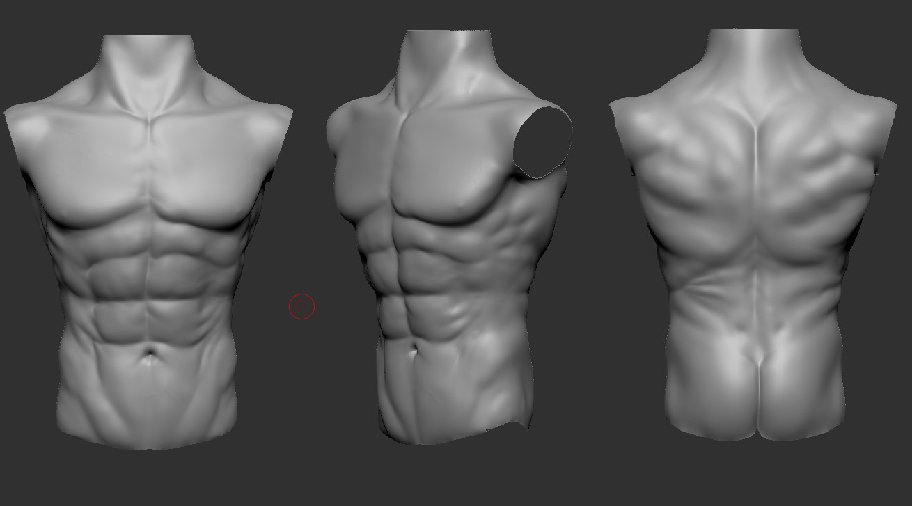
Artstation Male Chest Anatomy Practice Tomas Sosto from cdnb.artstation.com A line is drawn from anterior surface of the body of 6th thoracic vertebrae passing through the apex of the heart up to anterior lower most part of diaphragm. In insects, crustaceans, and the extinct trilobites, the thorax is one of the three main divisions of the creature's body, each of which is in turn composed of multiple segments. The clinical anatomy of the respiratory tract starts at the external nares or nose. Learn about each muscle, their locations & functional anatomy. This page provides an overview of the chest muscle group. It is enclosed by the ribs, the vertebral column, and the sternum, or breastbone, and is separated from the abdominal cavity (the body's largest hollow space) by a muscular and membranous partition, the diaphragm. The chest anatomy includes the pectoralis major, pectoralis minor and the serratus anterior. Learn about each of these muscles, their locations, functional anatomy and exercises for them.
The chest is the area of origin for many of the body's systems as it houses organs such as the heart, esophagus, trachea, lungs, and thoracic diaphragm.
The pig has a large horn plate, perforated by the two nares. It's also sometimes referred to as the breastbone. The right side of the heart is deflected anteriorly, and the left side is deflected posteriorly. The chest is made up primarily of two muscles: Anatomy of the breast, axilla, and chest wall. The chest anatomy includes the pectoralis major, pectoralis minor and the serratus anterior. Related posts of anatomy of the chest anatomy of stomach. Radiology basics of chest ct anatomy with annotated coronal images and scrollable axial images to help medical students and junior doctors learning anatomy. (1) the pectoralis major, and (2) the pectoralis minor. Of the two chest muscles, the pectoralis major (a.k.a. Related posts of anatomy of the chest area anatomy of the elbow. Anatomy of the thorax, heart, abdomen and pelvis recommended text gray's anatomy for students. The chest wall is comprised of skin, fat, muscles, and the thoracic skeleton.
Angina is the term for chest pain caused by poor blood flow to the heart. Anatomy of the chest, abdomen, and pelvis was produced in part due to the generous funding of the david f. Related posts of anatomy of the chest area anatomy of the elbow. Anatomy of the chest and stomach, human anatomy, anatomy of the chest and stomach. Milk line from the axilla to the groin.
Anatomy Chest Anatomy Drawing Diagram from i.pinimg.com The chest or thorax is the region between the neck and diaphragm that encloses organs, such as the heart, lungs, esophagus, trachea, and thoracic diaphragm. The epidermis is the outermost layer that provides a protective, waterproof seal over the body. Of the two chest muscles, the pectoralis major (a.k.a. It is not unusual to have areas of bruising or erosion to the dorsal tip of the rostal plane. The mammary bud grows downward into the dermis and starts branching to the secondary bud around the twelfth week. Your sternum is a bone that's located in the middle of your chest. Here, we break down the anatomy of your chest muscles. Anatomy of the thorax, heart, abdomen and pelvis recommended text gray's anatomy for students.
The chest anatomy includes the pectoralis major, pectoralis minor and the serratus anterior.
The chest is made up primarily of two muscles: Your sternum protects the organs of your torso from injury and also serves as a. Anatomy of the thorax, heart, abdomen and pelvis recommended text gray's anatomy for students. Anatomy of stomach 12 photos of the anatomy of stomach anatomy of gastric glands, anatomy of stomach and spleen, anatomy of stomach emedicine, anatomy of the stomach area female, parts of stomach ppt, human anatomy, anatomy of gastric glands, anatomy of stomach and spleen, anatomy of stomach emedicine, anatomy of the stomach area. About the 6th week, the somites differentiate into the sclerotomes and the dermatomyotomes. Related posts of anatomy of the chest anatomy of stomach. See human chest anatomy stock video clips. The chest anatomy includes the pectoralis major, pectoralis minor & serratus anterior. The pec major) is the one that commands the most real estate. The circulatory system does most of its work. Sternocleidomastoid muscle clavicle and ribs anatomy muscle anatomy chest sternocleidomastoid ribs anatomy chest muscles anatomy thorax rib muscles chest muscles chest anatomy illustration. Radiological anatomy of the lungs, mediastinal lymph nodes, trachea, bronchi, pleural cavity, heart and pulmonary vessels. Thoracic cavity, also called chest cavity, the second largest hollow space of the body.
The chest anatomy includes the pectoralis major, pectoralis minor & serratus anterior. The chest wall is comprised of skin, fat, muscles, and the thoracic skeleton. It's also sometimes referred to as the breastbone. Anatomy of the breast, axilla, and chest wall. Of the two chest muscles, the pectoralis major (a.k.a.
1 Anatomy Of The Arm And Chest Used With Permission Of N Moureau Download Scientific Diagram from www.researchgate.net The first step in understanding thorax anatomy is to find out its boundaries. Assoc prof craig hacking et al. The epidermis is the outermost layer that provides a protective, waterproof seal over the body. Radiological anatomy of the lungs, mediastinal lymph nodes, trachea, bronchi, pleural cavity, heart and pulmonary vessels. It is enclosed by the ribs, the vertebral column, and the sternum, or breastbone, and is separated from the abdominal cavity (the body's largest hollow space) by a muscular and membranous partition, the diaphragm. A heart attack results from blocked blood flow, often from a blood clot, to your heart muscle. The clinical anatomy of the respiratory tract starts at the external nares or nose. Here, we break down the anatomy of your chest muscles.
It is enclosed by the ribs, the vertebral column, and the sternum, or breastbone, and is separated from the abdominal cavity (the body's largest hollow space) by a muscular and membranous partition, the diaphragm.
Sternocleidomastoid muscle clavicle and ribs anatomy muscle anatomy chest sternocleidomastoid ribs anatomy chest muscles anatomy thorax rib muscles chest muscles chest anatomy illustration. Where is the sternum found. This article lists a series of labeled imaging anatomy cases by system and modality. The right side of the heart is deflected anteriorly, and the left side is deflected posteriorly. The mammary bud grows downward into the dermis and starts branching to the secondary bud around the twelfth week. 12 photos of the anatomy of the chest and stomach. Anatomy of the elbow 12 photos of the anatomy of the elbow anatomy and biomechanics of the elbow. Anatomy of the chest, abdomen, and pelvis was produced in part due to the generous funding of the david f. Here, we break down the anatomy of your chest muscles. Assoc prof craig hacking et al. Plus, how to target each to make them bigger and stronger. Milk line from the axilla to the groin. The epidermis is the outermost layer that provides a protective, waterproof seal over the body.
Share :
Post a Comment
for "Anatomy Of Chest / Muscles Of The Chest And Upper Back"


Post a Comment for "Anatomy Of Chest / Muscles Of The Chest And Upper Back"